
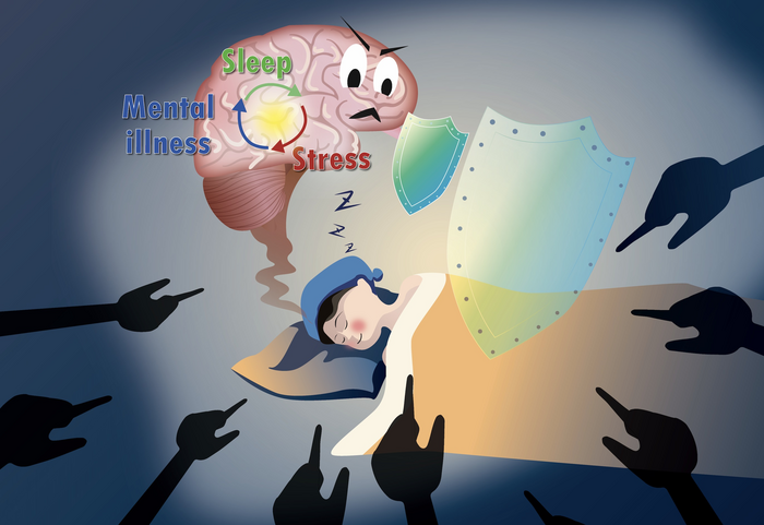

Stress promotes a kind of sleep in mice that subsequently relieves anxiety, according to new research that also pinpoints the mechanism responsible. The study, by scientists at Imperial College London, and colleagues across institutions in China, and at the University of Zurich, showed that in mice exposed to a type of psychosocial stress called social defeat stress (SDS), a subset of gamma-aminobutyric acid (GABA)-somatostatin neurons in the ventral tegmental area (VTA) of the brain receive and are activated by this stress input, promoting both REM (rapid-eye movement) and non-REM sleep, and also inhibiting the release of corticotropin-releasing factor (CRF). Together, the sleep initiated through this process alleviated stress levels and mitigated stress-induced anxiety in mice, restoring mental and body functions.
Since sleep is similar across mammals, it is likely the same mechanism is triggered in human brains. Uncovering the pathways involved could lead to the development of artificial ways to trigger these beneficial effects, potentially helping to treat persistent stress disorders such as PTSD, or to ease the psychosocial stress experienced by people who have been newly diagnosed with dementia.
Research lead Bill Wisden, PhD, at the Department of Life Sciences at Imperial, commented: “Our results add weight to the idea that REM [rapid eye movement] sleep helps us cope with stress. However, we previously only knew about ways REM sleep is reduced, such as some drugs that suppress it. Now, our study has revealed a mechanism by which REM sleep is induced, paving the way for drugs or other interventions that target the right neurons and boost the stress-busting power of sleep.”
Wisden and team reported on their studies and results in Science, in a paper titled “A specific circuit in the midbrain detects stress and induces restorative sleep.”
Humans and all mammals experience two main types of sleep. These are known as REM, when we tend to dream, and non-REM (NREM), which is deeper, dreamless sleep. While stress can cause insomnia and raise levels of stress hormones, the opposite can also be true, the authors noted. “Chronic stress increases rapid eye movement (REM) sleep; and sleep in rodents is induced by specific types of stress, such as social defeat stress (SDS).” Although the function and benefits of sleep remain unclear, sleep is certainly restorative, the team continued. “Thus, sleep has been suggested to be one of the mechanisms for alleviating the malign effects of stress.”
However, whether there is a specific circuit that links stress and sleep hasn’t been understood. The researchers reasoned that the ventral tegmental area of the midbrain could represent this link between stress and sleep. The VTA regulates reward, aversion, goal-directed behaviors, and social contact, they explained. “It also influences responses to stress and threats, and strongly affects sleep and wake … Because some γ-aminobutyric acid (GABA) VTA neurons are activated by stressful and aversive stimuli, we hypothesized that this route allows stress to induce sleep.”
For the reported study mice were exposed to a type of psychosocial stress called social defeat stress – which is used as an analogue for human bullying – by exposing them to particularly aggressive mice, without physical harm. The researchers found that after this encounter levels of flight or fight hormones in the SDS-exposed animals rose, indicating stress. When the mice then slept, the researchers then tracked the activity of the VTA neurons, revealing a specific subset of neurons that detected and responded to stress hormone levels and induced sleep high in both NREM and REM. “Activity-dependent tagging revealed a subset of ventral tegmental area γ -aminobutyric acid (GABA)–somatostatin (VTAVgat-Sst) cells that sense stress and drive non–rapid eye movement (NREM) and REM sleep through the lateral hypothalamus and also inhibit corticotropin-releasing factor (CRF) release in the paraventricular Hypothalamus,” the authors noted.
The activity of these neurons, and levels of NREM and REM sleep, stayed high for around five hours of sleep, during which signals were sent to other neurons that regulate stress hormones, blocking them from releasing more. The newly discovered neurons thus not only detected stress and induced sleep as a result, they also triggered the lowering of stress hormones.
The findings showed that sleep experienced by the mice appeared to lower the animals’ anxiety levels the next day. Once the mice awoke, the researchers tested their anxiety response to see how the sleep had affected their stress behaviors. This was evaluated by measuring how long the mice spent in the light, rather than seeking out darkness, as they will tend to do more when they are anxious. The animals’ responses were compared to those of stressed mice that were either sleep deprived (stimulated with objects) or had their newly identified neurons impaired, meaning that they didn’t get the same restorative sleep as did the normal mice.
The team found that mice that didn’t get their stress-induced sleep spent much more time in the dark, indicating that they were more anxious, and stress hormone levels also remained high in these animals. “For mice allowed sufficient home cage sleep after SDS, raised CORT [corticosterone] concentrations
returned to baseline over 60 min,” the authors noted. “If mild sleep deprivation occurred immediately after stress, however, CORT concentrations remained elevated … “For mice unable to have SDS-induced sleep, either because their VTASst neurons had been ablated or were inhibited, CORT concentrations remained higher during their home cage sleep after SDS, similar to the effects of sleep deprivation after SDS.”
Having identified this new mechanism, the team now hope to find ways to selectively target the responsible neurons and boost their positive effects via sleep. This strategy might be used to help treat persistent stress disorders such as post-traumatic stress disorder (PTSD). People who suffer from PTSD experience less REM sleep, contributing to the theory that REM sleep helps us process difficult emotions and stress.
Dementia diagnosis can also cause significant psychological stress, and the team hopes that if their research can lead to a way to boost the effects of sleep, this will also help people cope with a new diagnosis. People living with dementia in addition suffer from more emotional disturbances, and boosting REM sleep may similarly help reduce this distress. The authors concluded, “Thus, a specific circuit allows animals to restore mental and body functions by sleeping, potentially providing a refined route for treating anxiety disorders.”
In a perspective in the same issue of Science, Marian Joëls, PhD, at Utrecht University, and E. Ronald de Kloet, PhD, at Leiden University, noted that the reported results highlight a neurological circuit in the brain, the targeting of which “may help steer future interventions in rodents and perhaps even humans after stressful experiences, be it through cognitive therapy or pharmacotherapy or maybe, one day, genetic interference.”
Joëls and Kloet note that “Not all individuals may respond to social defeat with a bout of sleep,” and that recent studies suggest that social stress promotes sleep-like inactivity in mice, but with a large degree of variation. Also, in the reported study not all of the animals showed a strong increase in REM sleep duration. The authors say this individual variation “requires further investigation in larger groups of mice …” and concluded, “Knowing the essential steps in the brain may help steer future interventions in rodents and perhaps even humans after stressful experiences, be it through cognitive therapy or pharmacotherapy or maybe, one day, genetic interference.”